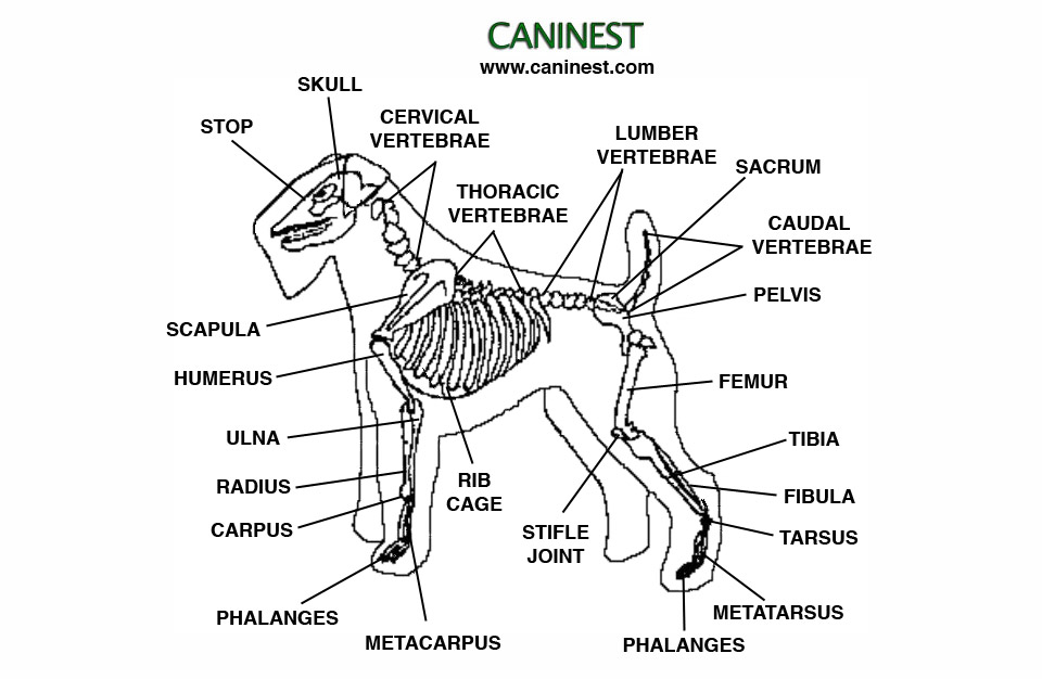
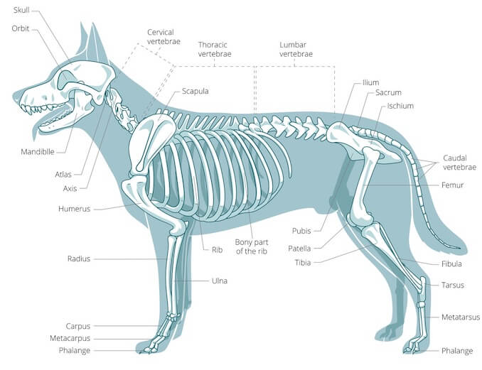
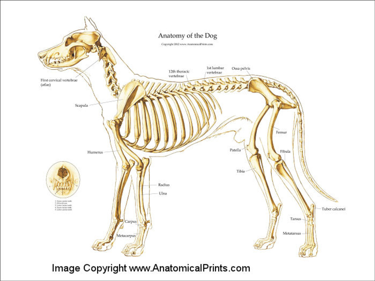
Dog Skeleton Anatomy
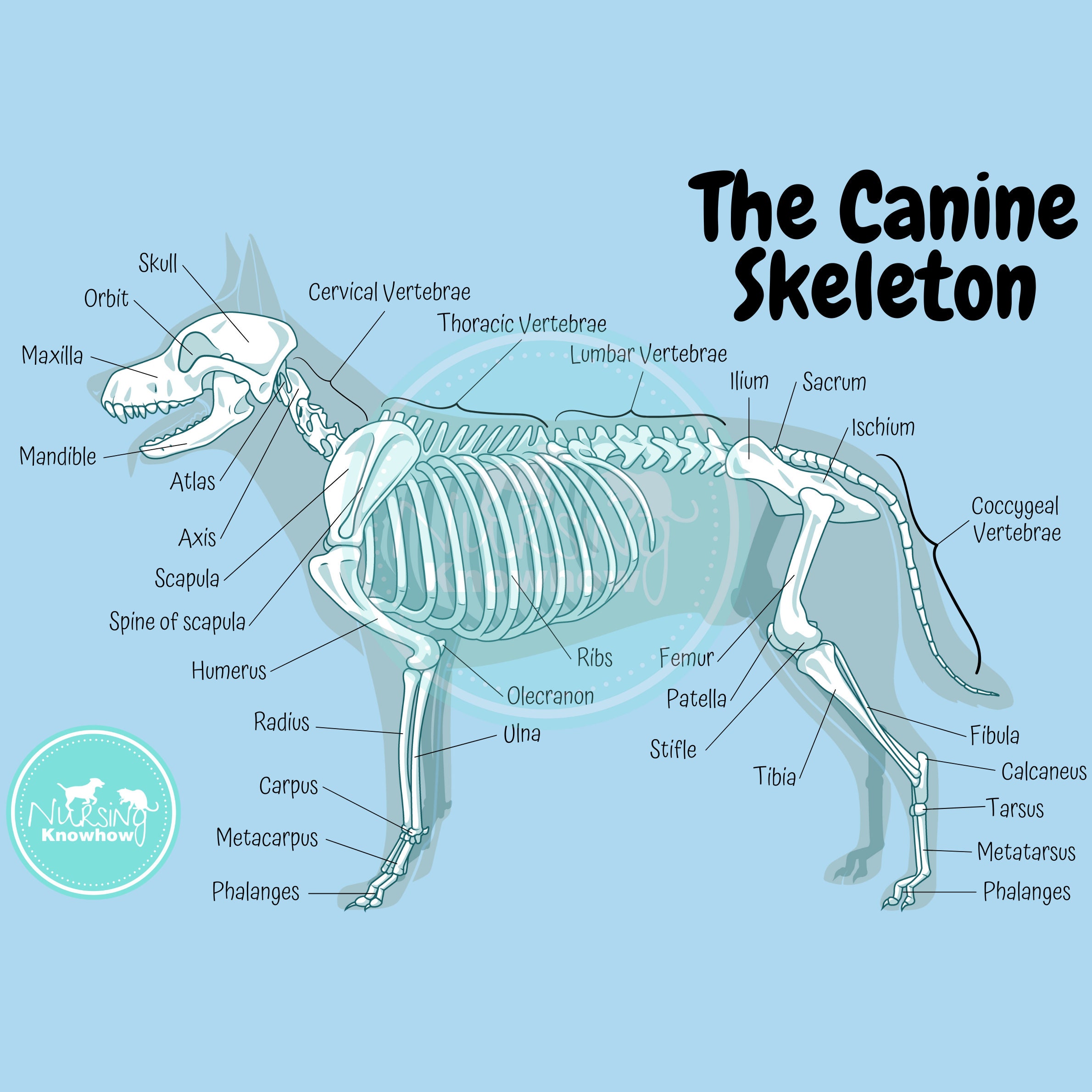
In the big picture, the dog skeleton is made of two basic parts: axial and appendicular (limbs). Axial skeleton = head (skull) + the spine (made of vertebrae) + ribs + sternum. Appendicular = forelimb bones + hindlimb bones. The axis of the dog skeleton is composed of the skull, spine (made of vertebrae), ribs and sternum (made of sternebrae).

Full dog anatomy with skeleton and internal organs, digital illustration. — computer
This veterinary anatomy module contains 608 illustrations on the canine myology. Here are presented scientific illustrations of the canine muscles and skeleton from different anatomical standard views (lateral, medial, cranial, caudal, dorsal, ventral / palmar.). Some fascias, tendons, ligaments, joints were labeled.
A Visual Guide to Dog Anatomy (Muscle, Organ & Skeletal Drawings) All Things Dogs
The dog skeleton anatomy consists of bones, cartilages, and ligaments. You will find two different parts of the dog skeleton - axial and appendicular. Here, I will show you all the bones from the axial and appendicular skeleton with their special osteological features. Again, I will provide more labeled diagrams for each dog skeleton bone.

FileDog anatomy lateral skeleton view.jpg
Dog anatomy comprises the anatomical studies of the visible parts of the body of a domestic dog.Details of structures vary tremendously from breed to breed, more than in any other animal species, wild or domesticated, as dogs are highly variable in height and weight. The smallest known adult dog was a Yorkshire Terrier that stood only 6.3 cm (2.5 in) at the shoulder, 9.5 cm (3.7 in) in length.

Dog skeleton with major bone elements labeled (Davis, 1987, p. 54;... Download Scientific Diagram
A dog's skeleton is made up of many different bones, which provide structure and support for their body. Dogs have over 300 bones in their body, which is more than humans who have around 206 bones. Their skeleton includes their skull, spine, ribcage and limbs. Dogs have four legs that are designed to help them move quickly and efficiently.

Labeled Dog Skeleton Diagram
This is an overview of the canine skeleton, pointing out features that help in identification and orientation.If you find this helpful, please let me know by.
Canine Skeleton Poster Clinical Charts and Supplies
Labeled anatomy of the head and skull of the dog on CT imaging (bones of cranium, brain, face, paranasal sinus, muscles of head) This module of vet-Anatomy presents an atlas of the anatomy of the head of the dog on a CT. Images are available in 3 different planes (transverse, sagittal and dorsal), with two kind of contrast (bone and soft tissues).
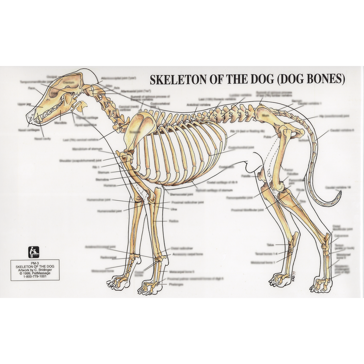
PetMassage™ Chart 3 Skeleton of the Dog · PetMassage™ Training and Research Institute
Summary Anatomy of a Dog Dog anatomy details the various structures of canines (e.g. muscle, organ and skeletal anatomy). The detailing of these structures changes based on dog breed due to the huge variation of size in dog breeds. Would you be surprised to know that short dogs are more aggressive? Or taller dogs are more affectionate?

Dog skeleton with major bone elements labeled (Davis, 1987, p. 54;... Download Scientific Diagram
How are dog skeletons adapted? Dogs have skeletons disconnected shoulder bones that are very similar to human skeletons. Their spine, skull, and limbs have adapted for their body weight by being sturdier than the same bones in people would be. They also have a light rib cage to run quickly without tiring out too easily.

Dog Anatomy Dog Skelton
The skeleton is the framework upon which the muscles, tendons, and ligaments are built to provide locomotion for the dog and to serve as a means of protecting the internal organs. For the most part, the composition of the skeleton dictates the movement of the dog.

Skeletal abnormality in dogs and cats
Components of the Musculoskeletal System in Dogs. Bones provide rigid structure to the body and shield internal organs from damage. They also house bone marrow, where blood cells are formed, and they maintain the body's reservoirs of calcium and phosphorus. Old bone tissue is constantly replaced with new bone tissue in a process called.

Dog skeleton 101 Dog Anatomy Bones Animal Hackers
The cat has a small coronoid fossa medial to the radial fossa that accommodates the coronoid process of the ulna during elbow joint flexion.; The cat has a supracondylar foramen near the medial condyle allowing the passage of the median nerve and brachial blood vessels.; There is an intermediate tubercle between the greater and lesser tubercles in the horse's intertubercular groove.

Anatomy of dog skeleton with labeled inner bone scheme vector illustration Dog skeleton
Dog skeleton Muscles of the dog Organs of dogs Canine anatomy As we explain above, canine anatomy is far ranging due to the diversity of existing breeds. These different breeds not only differ from each other in size, but in the shape of many body parts. Perhaps the most significant is head shape.
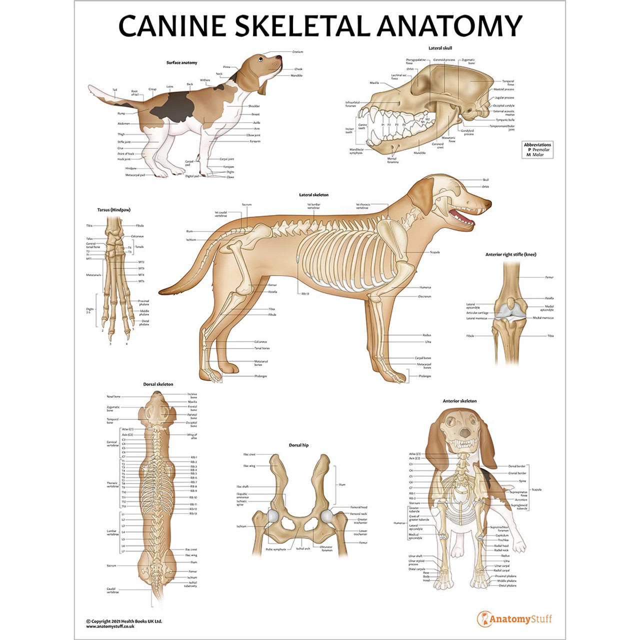
Canine Skeletal Anatomy Laminated Chart Dog Skeleton Poster
Our canine charts cover internal organ anatomy, the musculoskeletal system, common pathologies and guides to dog health and safety. Excellent wall displays in vet clinics, surgeries, dog groomers, and veterinary colleges. Our canine posters are s uitable for both animal lovers and veterinary studies.. Our canine model range covers detailed pathology models, common canine parasite models.
The Canine Skeletal Anatomy Poster Dyslexia Blue Etsy UK
An overview of the anatomy of the canine skeleton.Follow on twitter @ https://twitter.com/PerkyVetInstagram: Perkydvm

maryrosedog Dog skeleton, Dog anatomy, Dog skull
Anatomic Planes The main planes of motion for dogs are as follows (see Figure 5-1): • The sagittal plane divides the dog into right and left portions. If this plane were in the midline of the body, this is the median plane or median sagittal plane. • The dorsal plane divides the dog into ventral and dorsal portions.